From Cole's stepped tube to a pressure-controlled clinical trial, leakage, exchange, and laryngeal injury remained different questions.
Reading Library · Anesthesia Epochs · 中文版
Watch the companion film
1945–2009: Cuffed or Uncuffed? Pediatric Tracheal Tubes · Watch on YouTube
Historical anchor: 2009. Date details and source qualifications are explained below.
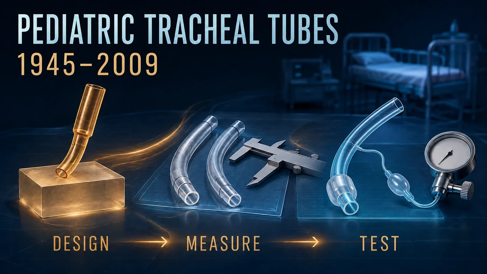
(Figure 1) Design, measurement, and testing. Original retrospective illustration, not to scale or a photograph of the original 1945 object. The modern tube's airway lumen and cuff inflation circuit are separate.[1–3]*
Two pairs of percentages from a 2009 trial tell different stories.
For tube exchange, the figures were 2.1% and 30.8%. For early post-extubation stridor, they were 4.4% and 4.7%. The first contrast was large and statistically significant; the second was not. Yet even before asking what those results mean, a reader needs to notice something less conspicuous: the two outcomes were reported using different grouping rules.[3]
That detail is a useful entrance to the history of pediatric tracheal tubes. A tube is not simply cuffed or uncuffed. Its outer diameter, cuff geometry, depth mark, inflation pressure, material, and method of use all affect what is being tested. Likewise, a successful seal, fewer exchanges, and an absence of detected early stridor are not interchangeable endpoints.
The path from Frank Cole's stepped tube in 1945 to the multicenter Microcuff trial in 2009 becomes more interesting when these distinctions remain visible. It is a story about making the question precise enough to investigate, not about adding a balloon and declaring the problem solved.
What the 2009 comparison actually tested
Weiss and colleagues organized a randomized trial across 24 European pediatric anesthesia centers, coordinated from Zurich. Its report appeared online on 3 November 2009.[3]
The cuffed intervention was a specific pediatric Microcuff tube used with a size-selection scheme, depth markings, a cuff manometer, and a pressure-release valve. Cuff pressure was limited to 20 cm H₂O under the study protocol. These conditions were not incidental accessories. They were part of the intervention whose performance the investigators observed.
The uncuffed group used the centers' usual tubes and institutional sizing practices. Before anesthesia induction, staff opened sealed, opaque, consecutively numbered allocation envelopes. If a tube did not fit or provide adequate ventilation, the protocol allowed exchange; some children consequently ended up with a different tube type from the one initially assigned.[3]
The population was selected, too. Participants were younger than five years, ASA physical status I or II, with a lower weight boundary around 3 kg, planned controlled ventilation, supine procedures, and intended extubation in the operating theater. Important exclusions included known airway abnormalities, anticipated difficult intubation, specified pulmonary disease, regurgitation or aspiration risk, and planned postoperative ICU ventilation. The trial was not a test of every neonatal airway or prolonged intensive-care intubation.[3]
Even the pressure measurements require care. An airway inflation-pressure leak test with the cuff fully deflated helped judge whether the tube was too large. Intracuff pressure was then adjusted to obtain a seal during ventilation. Those measurements shared a unit but not a purpose. Treating them as one universal “leak pressure” would obscure both the protocol and the device's behavior.
Fewer exchanges, without a detected stridor difference
The centers returned 2,406 data forms. Investigators excluded 160 because the age group or age-related tube size had not been selected correctly, leaving 2,246 children for analysis: 1,119 in the initially cuffed group and 1,127 in the initially uncuffed group.[3]
By initial allocation, 24 cuffed-group children required a tube exchange, compared with 347 uncuffed-group children: 2.1% versus 30.8%, P<0.0001. These are patients who underwent exchange, not the total number of exchange procedures, which was higher because some children needed more than one.
The reported stridor comparison was different. It classified children primarily by the final tube used, after exchanges, and found rates of 4.4% with cuffed tubes and 4.7% with uncuffed tubes, P=0.543. It was therefore not simply the same initial-allocation comparison applied to a second outcome. The source tables also retain final-group labels that do not fully reconcile with the text's exclusion of five children from morbidity assessment. The reported percentages are preserved here; no corrected denominator has been invented.[3]
An independent assessor recorded new high-pitched inspiratory sounds within one hour after extubation, with attention to pain, secretions, and residual airway obstruction. That is a clinically meaningful early observation. It is not systematic endoscopic inspection, a measure of every mucosal lesion, or long-term follow-up for subglottic stenosis.
The defensible result is consequently specific: the tested approach markedly reduced the need for tube exchange, while the study did not detect a statistically significant difference in early stridor. Non-significance does not establish equivalence, and neither endpoint establishes universal freedom from airway injury.
Why geometry had to come before the trial
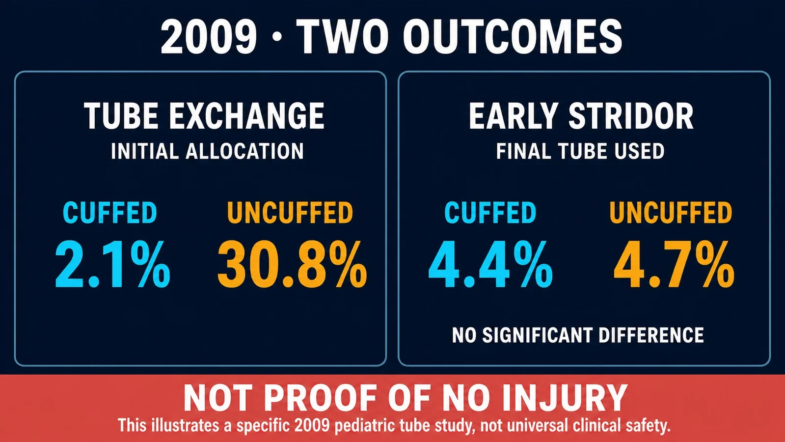
(Figure 2) Two outcomes, two grouping rules. Exchange rates of 2.1%/30.8% use initial allocation; early stridor rates of 4.4%/4.7% use the final tube. No detected significant stridor difference is not proof of no injury. The panels do not share a denominator.[3]*
To understand why the study combined a tube with sizing, depth marks, and pressure control, it helps to step back to an earlier investigation by Weiss, Dullenkopf, Gysin, Dillier, and Gerber.
They obtained tube samples in 2002; the resulting paper was accepted in 2003 and published in the January 2004 issue of the British Journal of Anaesthesia. It examined 125 tubes from four manufacturers across 15 product series, 11 cuffed and four uncuffed. The investigators measured outer diameter, cuff dimensions and location, and depth markings, then compared those measurements with age-related anatomical reference data.[2]
Nominal internal diameter did not settle external fit. Tubes carrying the same internal-diameter size could have different outer diameters. The lumen determines one aspect of airflow, but the shaft wall and cuff contribute to the structure placed against the airway. A number printed on a tube cannot stand in for all of those dimensions.
Length created a second difficulty. With the tip positioned at the reference mid-tracheal level, some cuffs would occupy the laryngeal region. Advancing the tube to move the cuff below that region could bring its tip too close to the carina. Improving clearance at one end could consume the margin at the other.[2]
Only five of the 11 cuffed series had depth marks, and existing marks did not consistently provide the desired placement margin. The authors argued for a short high-volume, low-pressure cuff, a cuff-free subglottic segment, and appropriately located depth markings. These were connected design requirements, not independent decorative features.
The study made a geometric problem visible. It did not measure clinical injury rates, inspect every child directly, or encompass all products, preformed tubes, and manufacturing lots. Its older anatomical assumptions and recommendations should not be repackaged as present-day bedside instructions. Its enduring historical contribution is narrower: a debate framed only as cuffed versus uncuffed had overlooked the shapes being compared.
Cole's earlier answer to a different constraint
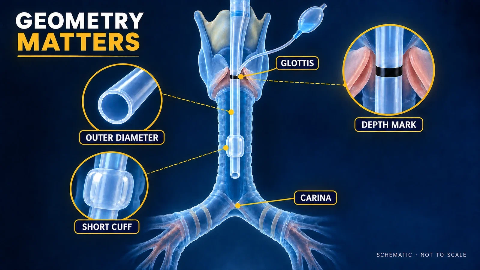
(Figure 3) Geometry matters. A short cuff lies below the subglottic region, with space between tube tip and carina. This not-to-scale design schematic specifies no clinical insertion depth or safe distance.[2]*
The Wood Library-Museum dates Cole's pediatric tube to 1945, when endotracheal anesthesia in infants and young children was still uncommon. His solution addressed the resistance imposed by a narrow tube.[1]
The patient-end segment was narrow enough for the confined passage, while the upper shaft was wider. The entire length therefore did not have to share the narrowest section's dimensions. The museum also describes the wider portion as helping limit excessive insertion. This is a historical design rationale, not a quantified clinical benefit inferred from a paper not covered here.
Cole's 1957 publication on pediatric formulas belongs to a later stage. The museum catalog distinguishes the January 1945 paper, A New Endotracheal Tube for Infants, from the November 1945 paper, An Endotracheal Tube for Babies. Similar titles do not make them a single report.[1]
The catalog carries another warning for visual history. Its photographed clear-plastic Cole-type tube is dated approximately 1989, with the cataloger explicitly preserving uncertainty. It illustrates the stepped design but is not the original 1945 object. Using the later specimen as evidence of the design is reasonable; presenting it as an artifact photographed at the moment of invention would not be.
A chronology that must not collapse
The cuffed-versus-uncuffed clinical comparison by Khine and colleagues was published in 1997. Dullenkopf, Gerber, and Weiss's 2003 Microcuff paper was an in-vitro investigation. The large pediatric multicenter randomized report discussed here was published in 2009.[3–5]
These are distinct studies with distinct evidentiary roles. A laboratory test of fluid leakage cannot be renamed a pediatric randomized trial, and a change in cuff material cannot by itself establish prevention of clinical aspiration.
For this article, the complete 2004 design paper and 2009 trial were read. The full 1997 and 2003 papers are not covered. Their authoritative bibliographic records and abstracts establish identity and study type here, but their exclusive numerical results are not used. Preserving that distinction is more valuable than presenting a longer reference list as if every source had been read equally deeply.
The material change that interrupted the experiment
The 2009 paper contains a complication that a simple progress narrative could easily discard. The initial sample-size calculation called for 3,928 participants. A manufacturer change in the tube shaft material then increased the risk of kinking and led to a temporary recall. Correcting the problem required months, and the study stopped ahead of schedule. The investigators reported that an improved version became available about a year after the stop.[3]
The interruption belongs in the history, not outside it. A device's material is part of its performance, just as its dimensions and pressure management are. The trial did not reach its planned size, and the recall makes it especially inappropriate to detach a reassuring outcome from the particular device version and conditions that produced the evidence.
The papers also disclose equipment supplied by manufacturers and investigators' development relationships. Those disclosures do not automatically invalidate the results. They show the setting in which device development and clinical evaluation met.[2,3]
Progress with the endpoints left intact

(Figure 4) Five distinct steps. Tubes, bench apparatus, caliper, hospital network, and gauge are symbolic illustrations, not reconstructions of study apparatus. Recall and early stopping remain a side branch; the open track is not a universal-safety verdict.[1–5]*
Cole's design addressed resistance. The 2004 measurements exposed a conflict between fit, cuff position, and tip clearance. The 2009 trial examined a specific solution under defined clinical conditions. None of those kinds of evidence could do the work of all the others.
The historical interpretation offered here is that pediatric tube design became a more explicitly measured system: geometry, anatomical margins, cuff-pressure management, manufacturing behavior, and clinical outcomes were considered together. That interpretation does not require a claim that one product ended the debate.
Even a successful laboratory fluid seal would not demonstrate that patients no longer aspirate. A similar rate of early stridor does not demonstrate the absence of long-term injury. Keeping those limits in view does not diminish the observed reduction in tube exchange. It tells us exactly which advance the evidence can support.
Sources and reading limits
- Wood Library-Museum of Anesthesiology. Cole Tracheal Tube: description and complete catalog · Archived copy. Read, including specimen dating. The original Cole papers were identified, not represented as full-text read.
- Weiss M, Dullenkopf A, Gysin C, Dillier CM, Gerber AC. Shortcomings of cuffed paediatric tracheal tubes. Br J Anaesth. 2004;92:78–88. Complete published-version PDF from the University of Zurich repository read and visually inspected.
- Weiss M, Dullenkopf A, Fischer JE, Keller C, Gerber AC; European Paediatric Endotracheal Intubation Study Group. Prospective randomized controlled multi-centre trial of cuffed or uncuffed endotracheal tubes in small children. Br J Anaesth. 2009;103:867–873. Complete official PDF read and inspected.
- Khine HH et al. PubMed record of the 1997 clinical comparison. Identity and study-type use only; complete paper not covered.
- Dullenkopf A, Gerber A, Weiss M. Publisher record of the 2003 in-vitro Microcuff study. Identity/design use only; complete paper not covered, no exclusive numerical results used.
Anesthesia Epochs · 1945–2009.
Original editorial schematics are not to scale and are neither photographs of original historical objects nor clinical instructions.
Continue exploring
Related reading connects shared questions and contexts; it does not by itself establish historical influence.
- 1878: Macewen's Oral Tracheal Tube
- 1953: Virginia Apgar Gives the Newborn a Common Language
- 2013: An Old Airway Algorithm Makes Room for Video Laryngoscopy
- 2016: 1,018 Difficult Intubations Make 'Try Again' a Measurable Risk
- 2016–2019: GAS, PANDA and MASK Narrow the Pediatric Neurotoxicity Question
The Source Room · About & Editorial Method
Sources and reading limits are stated above. Illustrations are labelled as original source images or AI-made. Historical accounts are educational; they are not current treatment instructions.