In June 1993 the Apparatus section of Anaesthesia (vol. 48, no. 6) printed 'The levering laryngoscope' by E. P. McCoy and R. K. Mirakhur of the Department of Anaesthetics, The Queen's University of Belfast, accepted on 26 January 1993. Forward displacement of the larynx, prominent upper teeth and backward displacement of the tongue, they wrote, make intubation difficult; the force needed to lift the epiglottis then rises, and a curved blade is often levered on the upper incisors, which may be damaged. Earlier modifications had left the fulcrum near the teeth rather than at the epiglottis, and blades could not change shape once inserted. Their blade differed from the curved blade in four respects: a hinged tip, a lever at the proximal end, a spring-loaded drum and a connecting shaft. Pressing the lever with the thumb raised the tip, lifting the hyo-epiglottic ligament and exposing the larynx; releasing it returned the tip. The authors argued that moving the fulcrum towards the epiglottis would reduce force, could turn a grade 2 or 3 view into grade 1 or 2 without an assistant's laryngeal pressure, and might lessen the pressor response. The report is a device description with no clinical series; Penlon helped develop the blade and a patent was pending.
Reading Library · Anesthesia Epochs · 中文版
Watch the education video (for clinicians)
1993: The McCoy Levering Laryngoscope — Move the Fulcrum · Anesthesia Epochs Education Series
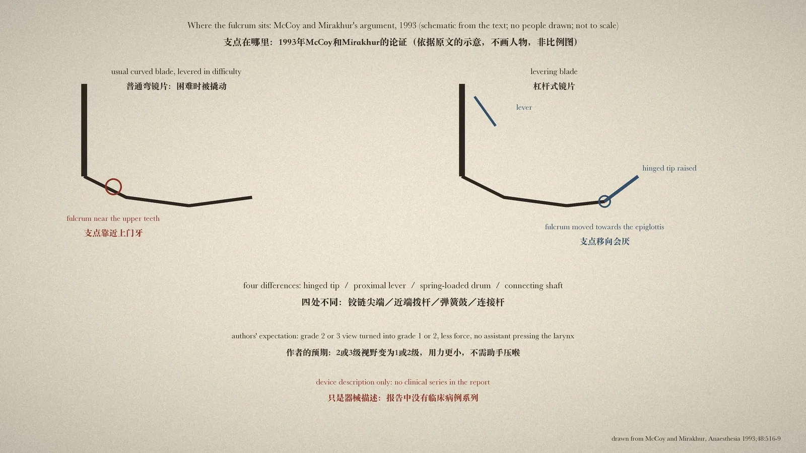
When the laryngoscope shows the epiglottis but not the glottis, the operator may start, almost without thinking, to rock the handle back and use the upper incisors as a fulcrum. Teeth get damaged and the view may not improve. In 1993 two anaesthetists in Belfast offered a different idea in a short device report: if the fulcrum is in the wrong place, let the blade change its shape during laryngoscopy. [1, p. 516]
Why the fulcrum ends up on the teeth
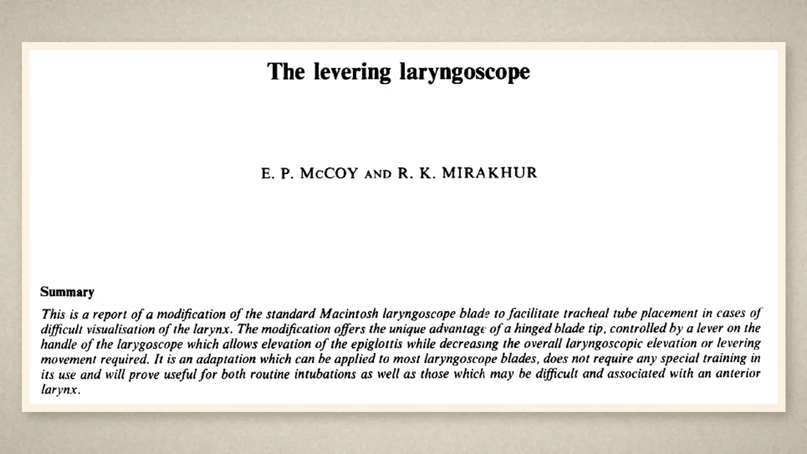
In June 1993 the Apparatus section of Anaesthesia (vol. 48, no. 6) printed "The levering laryngoscope" by E. P. McCoy, senior registrar and senior tutor, and R. K. Mirakhur, senior lecturer, of the Department of Anaesthetics at The Queen's University of Belfast. It had been accepted on 26 January 1993. [1, p. 516]
The introduction drew on Cormack and Lehane: the three main causes of difficult intubation are forward displacement of the larynx, prominent upper teeth and backward displacement of the tongue. The epiglottis can usually be exposed, but with limited mouth opening, a large tongue, a receding mandible, protruding teeth or a fixed cervical spine, lifting it may be difficult or impossible, and the force applied rises with the difficulty. With the curved Macintosh blade the force should run along the axis of the handle, but in difficult cases the blade may be levered, the upper teeth may "inadvertantly be used as a fulcrum", and persistent attempts to lift the epiglottis often damage them. A blade that avoided the incisors and had its fulcrum lower in the pharynx, the authors suggested, might make the epiglottis easier to lift. [1, p. 516]
Four changes
The new blade differed from the usual curved blade in four respects: a hinged tip, a lever at the proximal end, a spring-loaded drum and a connecting shaft. The blade was cut near its tip and a hinge put in; the flange was cut in a curve so that, at rest, the tip locks with the rest of the blade and pressure on the tip passes along the flange rather than through the hinge. The lever, at the proximal end, connects through a pin to a spring-loaded drum enclosed on the left side of the flange, and a connecting shaft links the drum to the hinged tip. [1, p. 516][1, p. 517]
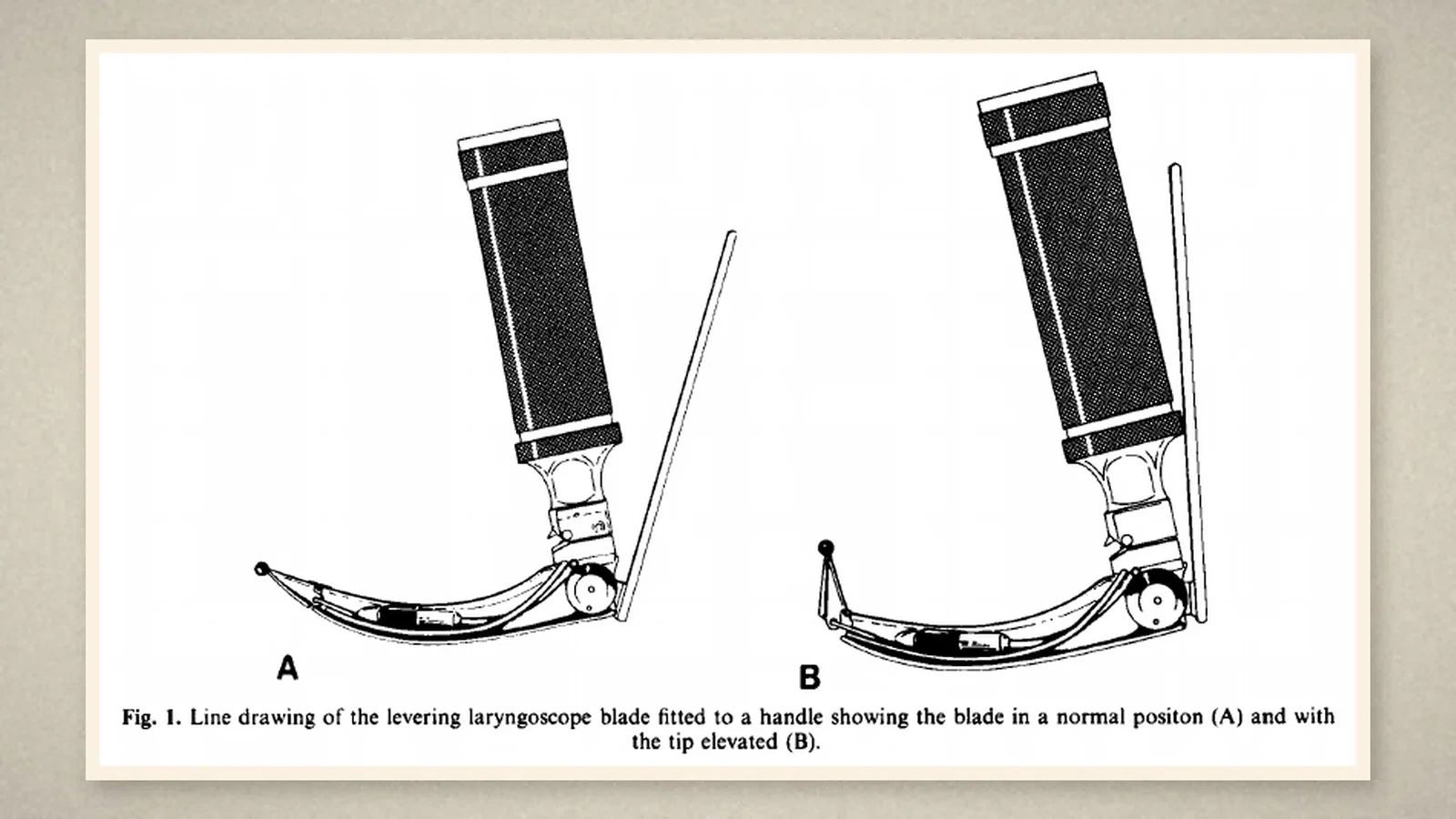
The authors admitted that the modified blade was somewhat heavier than an ordinary one, but judged this insignificant beside the weight of the handle and batteries. It fitted a standard Penlon handle. [1, p. 517]
A press of the thumb
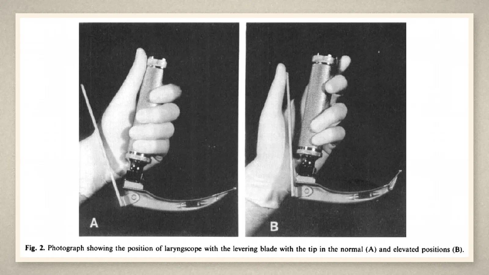
The handle is held in the normal way, with the lever lying behind the thumb. Pressing the lever towards the handle rotates the drum, pushes the connecting shaft forward along the blade and raises the tip; releasing it lets the spring return the tip to rest. [1, p. 517] Laryngoscopy proceeds as usual: the tip goes into the vallecula, the operator moves the thumb behind the lever and presses gently, and the tip rises, "lifting the hyo-epiglottic ligament and exposing the larynx". After the tube is placed the lever is released, the blade returns to its usual shape and is withdrawn normally. [1, p. 518]
Earlier attempts and the authors' expectations
The discussion listed earlier modifications: Orr's two right-angled bends, which placed the fulcrum within the oropharynx; a more concave midsection; a wider angle between blade and handle; an adjustable adaptor on the handle; an angulated blade with a prism to see round the corner; a lowered flange; a straight-shafted instrument with a curved tip; and a tip made adjustable by a screw-lock joint. Even so, the authors argued, the fulcrum stayed near the upper teeth rather than at the epiglottis, where it would be most effective; existing blades, "once inserted, are inflexible", and any movement of the tip at the epiglottis needed a much larger movement of the handle. The new blade moved the fulcrum closer to the area to be seen. [1, p. 518]
Their reasoning ran on. In grade 1 and 2 views less force should be needed, because the tongue need only be pushed aside while the hinged tip lifts the epiglottis; in grade 3, where the blade is often levered at the mouth, most of that force would be unnecessary if the fulcrum moved towards the epiglottis. Using the lever could "convert a grade 2 or 3 into a grade 1 or 2 intubation" without an assistant pressing on the larynx, and the tip could also be placed behind the epiglottis like a straight blade. In routine use, by reducing the force on the supraglottic tissues, it might also reduce the sympatho-adrenal response to laryngoscopy. The conclusion called it a "unique modification" allowing the blade to change shape during laryngoscopy and reducing the force needed to see the larynx. [1, p. 518]
The acknowledgements thanked three people at Penlon (UK) Ltd for help in developing the blade and stated that it had been patented pending further development and marketing. [1, p. 518]
What the record shows
The report rests on a mechanical judgement: in difficult laryngoscopy the trouble is not only that the larynx cannot be seen but that the fulcrum is in the wrong place. What it offers is a device and an argument. It has no patients and no force measurements, and its benefits are, in the paper itself, still the authors' expectations.
Sources and reading limits
Read: McCoy and Mirakhur, "The levering laryngoscope", Anaesthesia 1993;48:516-9 (the whole report: summary, introduction, description of the blade, use, discussion, acknowledgements and Figs 1 and 2). Obtained through institutional access.
Not covered: the force studies (Bucx and colleagues) and earlier blade modifications (Orr, 1969, and others) that the authors cite; clinical trials of the blade; the maker's records on its marketing. The History of Anaesthesia Society timeline entry was used only as a lead.
Limits: this is a device description with no clinical series, no measured forces with the new blade and no comparison with other blades. Converting grade 2 or 3 views into grade 1 or 2 and reducing the pressor response are the authors' expectations, not results. Penlon helped develop the blade and a patent was pending. Blade dimensions, weights and angles are not repeated. Fig. 2 shows only a gloved hand holding the laryngoscope, with no patient. "Unique" is the authors' word.
Three illustrations are crops of Anaesthesia 1993 pp. 516 and 517 (title and summary, the Fig. 1 line drawing, the Fig. 2 photographs). One is a schematic of the fulcrum argument drawn from the text, not to scale, with no people drawn.
- McCoy EP, Mirakhur RK. The levering laryngoscope. Anaesthesia. 1993;48:516-9. DOI
Continue exploring
Related reading connects shared questions and contexts; it does not by itself establish historical influence.
- 1984: Cormack and Lehane Grade the Laryngoscopic View in Four Levels
- 1985: Mallampati and Colleagues Propose a Pharyngeal Sign Predicting Difficult Intubation
Anesthesia Epochs · The Source Room · About & Editorial Method
Published November 3, 2026. Sources and reading limits are stated above. Illustrations are labelled as original source images or AI-made. Historical accounts are educational; they are not current treatment instructions.