Gordh's rubber-diaphragm needle and the Mayo Clinic's plastic needle, in their designers' own papers.
Reading Library · Anesthesia Epochs · 中文版
Watch the education video (for clinicians)
1945 and 1950: Gordh and Massa Needles — Staying in the Vein · Anesthesia Epochs Education Series
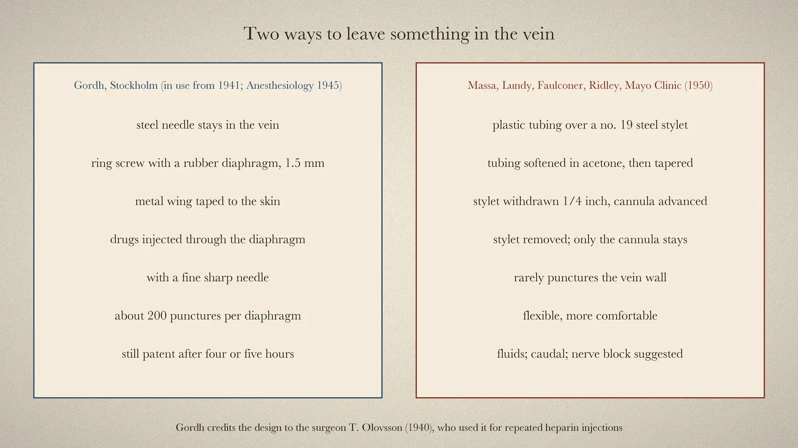
A plastic cannula left in a vein, through which drugs are given for hours, is now a familiar part of anaesthesia. Two short papers show earlier answers to the same problem: how to leave something in a vein that stays open, does not damage the vessel and lets the anaesthetist give repeated doses. One came from Stockholm in 1945, the other from the Mayo Clinic in 1950.
Stockholm: a needle with a rubber window
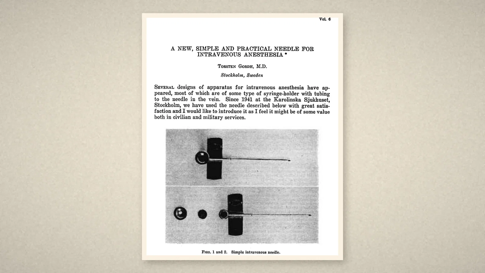
In May 1945 Torsten Gordh, of the Department of Anesthesia at the Karolinska Sjukhuset in Stockholm, wrote in Anesthesiology that several designs for intravenous anaesthesia had appeared, most of them a syringe-holder with tubing to the needle in the vein. Since 1941 Gordh's department had used something simpler, which Gordh thought might be of value in civilian and military services. [1, p. 258]
The needle, shown in actual size in the paper's first two figures, had a ring screw at a right angle to the shaft at its base. Inside the ring sat a rubber diaphragm 1.5 mm thick, and a metal wing at the base allowed it to be fixed to the skin with adhesive tape. [1, p. 258] The design was not Gordh's own: it had been made by a surgeon in Stockholm, T. Olovsson, described in Der Chirurg in 1940, for repeated injections of heparin. After some simple modifications the Karolinska department had adopted it for intravenous anaesthesia, and after three years they were still very satisfied with it. [1, p. 258–259]
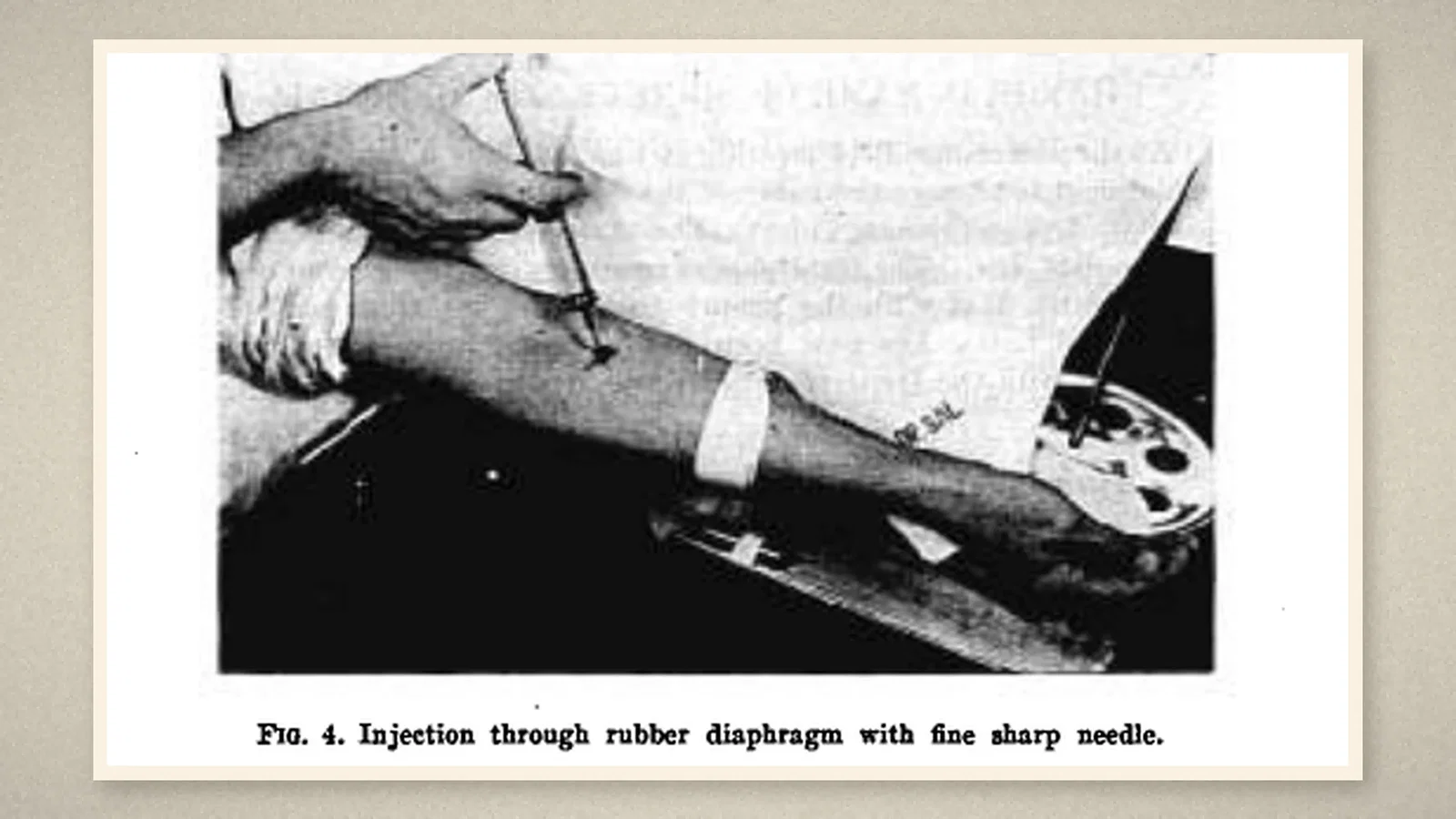
The venipuncture could be made wherever convenient: the cubital vein, the back of the hand or the foot. Movements of the arm did not spoil the arrangement as they could when a syringe was attached, and the patient could be moved from bed or stretcher to the operating table with the needle in place. [1, p. 259] The anaesthetic was then given with a very fine needle on the syringe, pushed through the rubber diaphragm at intervals; between injections the syringe lay on the arm-board. There was no need to aspirate blood into the syringe, clotting was seldom seen, and even after four or five hours of continuous use the passage remained patent. One diaphragm could take about 200 punctures and could be replaced at any time. The needle could also be used for a slow drip or a blood transfusion during the operation. [1, p. 260]
Rochester: a plastic sleeve over the needle
On 5 July 1950 the Proceedings of the Staff Meetings of the Mayo Clinic carried "A Plastic Needle" by David J. Massa, a fellow in anesthesiology, with John S. Lundy, Albert Faulconer Jr. and Roger W. Ridley of the Section on Anesthesiology. [2, p. 413–414] The needle consisted of a short length of transflex tubing fixed at one end to a hub and tapered at the free end, "through which a steel needle has been passed as a stylet". [2, p. 413]
The paper gave the recipe. The stylet was a 3-inch no. 19 Becton-Dickinson needle; the hub was a shortened no. 16 needle, notched so the tubing would grip it. Three-inch lengths of no. 22 tubing were soaked in acetone for two minutes, slipped over the stylet, dried, dipped again at the tip, and autoclaved; a smooth taper was then put on the tubing with a rotary cloth buffer so that its edge met the bevel of the needle. Assembled needles could be bought from the Rochester Products Company. [2, p. 413] The finished needle was sterilised by autoclaving for twenty minutes at 15 pounds' pressure. [2, p. 414]
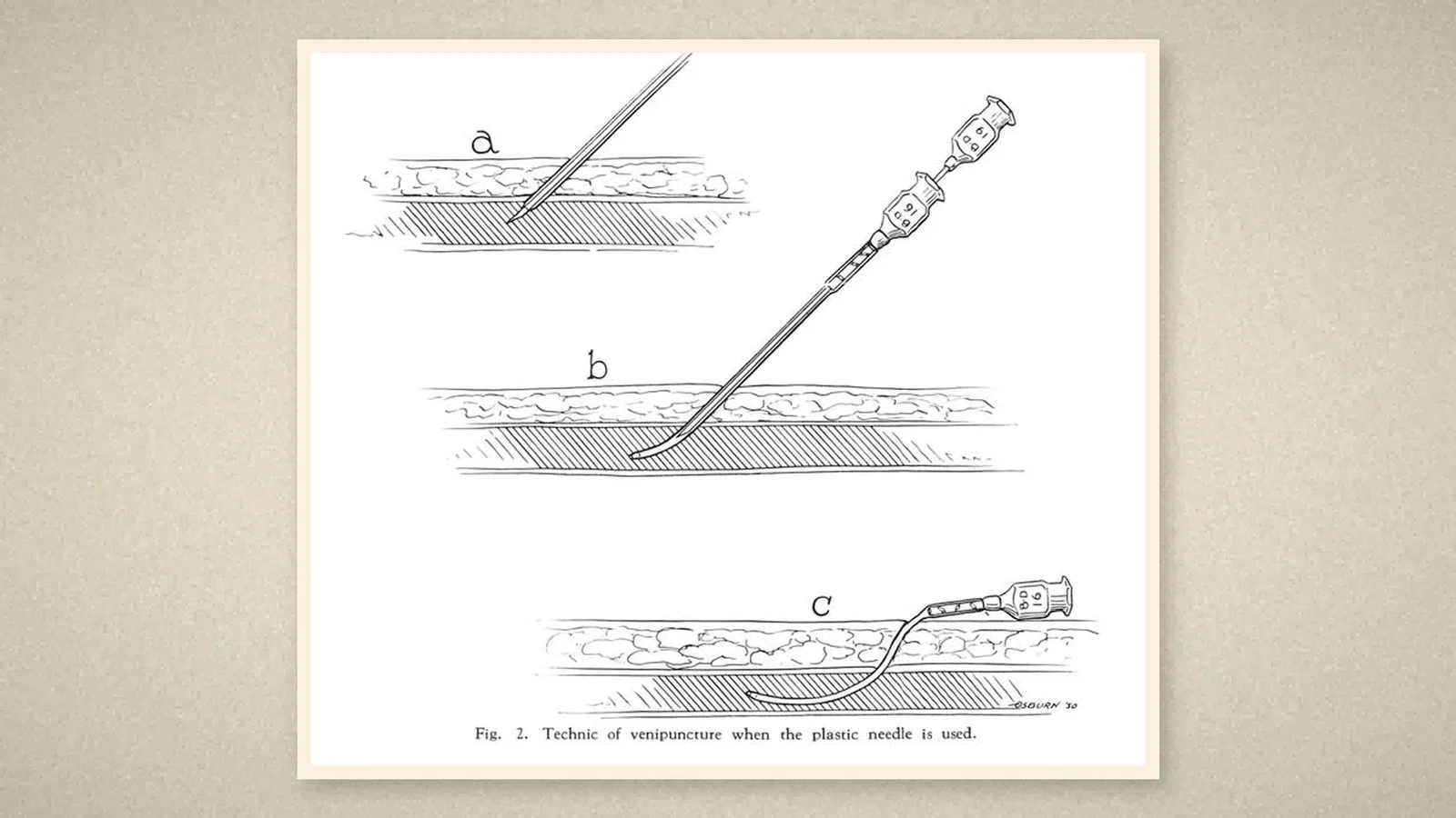
It was introduced into a vein like any needle. Once the tip of the cannula was well inside, the stylet was withdrawn about a quarter of an inch and the cannula was advanced as far as desired; then the stylet was removed entirely and fluid was connected to the hub, which was taped in the usual way. [2, p. 414] The authors gave three advantages: the cannula "will rarely puncture the wall of the vein as does a steel needle", its flexibility made it more comfortable for the patient, and putting it in was simpler than threading a plastic tube through a steel needle. [2, p. 414] It had proved most useful in the prolonged intravenous administration of fluids; a modification had been used in caudal anaesthesia, and continuous nerve blocks were suggested. [2, p. 415]
Two ideas
Gordh's needle left steel in the vein and solved the problem at the hub: a rubber window that could be punctured again and again, so that the syringe did not have to stay attached. The Mayo needle solved it at the tip: once in, the steel came out and only soft plastic remained. Gordh's paper was written for intravenous anaesthesia; the Mayo paper's main use was fluids over long periods. Neither gave numbers of patients or complications.
Sources and reading limits
Read: Gordh, Anesthesiology 1945, pp. 258–260, and Massa, Lundy, Faulconer and Ridley, Proc Staff Meet Mayo Clin 1950, pp. 413–415, both in full.
Not covered: Olovsson's 1940 paper in Der Chirurg was not read, so the needle's original form is known only from Gordh's sentence. Later developments, including a step sometimes dated to 1962, were not researched, and this article stops in 1950. No claim is made about which design came first anywhere else, or about later commercial cannulas.
One illustration is drawn from the two papers; three are crops of their pages.
- Gordh T. A new, simple and practical needle for intravenous anesthesia. Anesthesiology. 1945;6(3):258–260. doi:10.1097/00000542-194505000-00005
- Massa DJ, Lundy JS, Faulconer A Jr, Ridley RW. A plastic needle. Proc Staff Meet Mayo Clin. 1950;25(14):413–415. doi:10.1016/s0025-6196(26)04467-8
Continue exploring
Related reading connects shared questions and contexts; it does not by itself establish historical influence.
- 1909: Burkhardt Puts Ether into the Vein
- 1918: Howell and Holt Name the Liver's Anticoagulant Heparin
- 1934: Wisconsin Tries Abbott's Drug No. 8064 in 45 Patients
Anesthesia Epochs · The Source Room · About & Editorial Method
Published November 2, 2026. Sources and reading limits are stated above. Illustrations are labelled as original source images or AI-made. Historical accounts are educational; they are not current treatment instructions.